Radiation-Free Quantification of Intracranial Volume and Head Malformations from 3D Photography
Need:
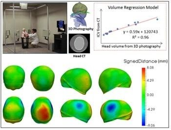
The evaluation of brain development in children with cranial pathology plays an essential role in surgical treatment and patient monitoring, in which the intracranial volume (ICV) and cranial malformations are two important measurements.
Solution:
We have developed a computational framework to quantify ICV and head malformations from 3D photography, and use them to characterize cranial shape abnormalities objectively and quantitatively in patients. A set of landmarks were automatically identified in 3D photographs of patients. That allows the cranial shape to be extracted and used to compute the head volume, cranial bone volume, intracranial/brain volume and cranial bone malformations at every location on the cranium.
Impact:
The novel automatic framework has the potential to reduce the use of radiation- and sedation-based imaging for surgical planning and patient monitoring. We are working with collaborators to collect more data and validate our framework. We are also working on extensions to support facial dysmorphology analysis from 3D photography.
Partners:
Funding:
NIH R42HD081712