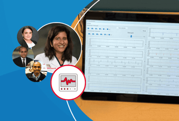
The Children’s National Hospital Sheikh Zayed Institute for Pediatric Surgical Innovation (SZI) is transforming what is possible to improve the technology innovation for pediatric device development. Anita Krishnan, MD, a pediatric cardiologist and clinician scientist, has research interests in advancing technologies for fetal cardiac diagnosis and improving access to early fetal diagnosis. Dr. Krishnan is a professor of pediatrics and a faculty member at the Sheikh Zayed Institute for Pediatric Surgical Innovation and Center for Prenatal, Maternal and Neonatal Health Research.
Dr. Krishnan’s research focuses on improving methods to diagnose and treat fetal heart problems in the very tiniest babies before they are born. Fetal arrhythmias are a condition in which a baby’s heartbeat is too fast or too slow during pregnancy. Within a day or two, without treatment, these conditions can progress quickly to heart failure and cause stillbirth. With early diagnosis and the right medications, babies can live a long and high-quality life and often outgrow them in the first year of life. In areas that are remote from the hospital, a baby may pass without anyone ever knowing the cause. Fetal heart problems can also lead to premature delivery. In some cases, the doctor may require the mom to take high risk cardiac medications during her pregnancy. This sometimes means long and complicated inpatient stays. Better home-based monitors could significantly reduce the inpatient stays for families.
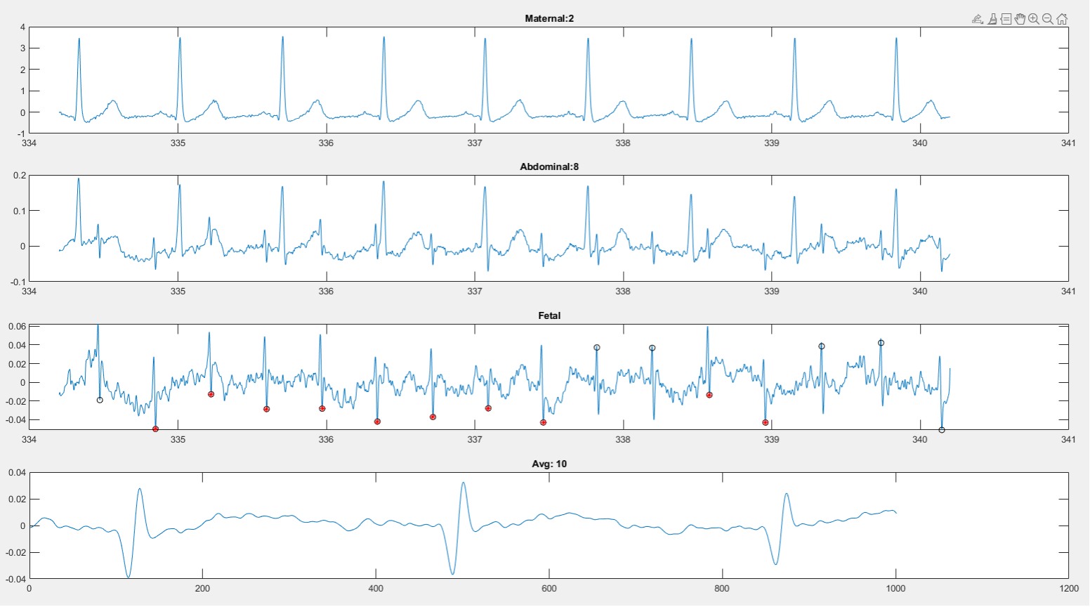
Dr. Krishnan and her team, Rathinaswamy Govindan, PhD, Pooneh Roshanitabrizi, PhD and Raj Shekhar, PhD, in collaboration with Auscultech Dx developed a novel technology to find the baby’s heart signal using a beat-to-beat fetal electrocardiogram. This innovation uses the differences in frequencies of the heart signal rather than the timing of the heartbeats. It works better at finding “beat-to-beat” separation where individual heart beats and more subtle arrhythmias become visible. The team has shown that the method is very accurate using their own standard and “gold standard” to synthesize data. In addition, it is well suited for situations where a baby is moving. In the last year Dr. Krishnan and the Auscultech team have made a device which is much smaller with the goal that it can eventually be portable at low cost for the uses in rural and remote regions.
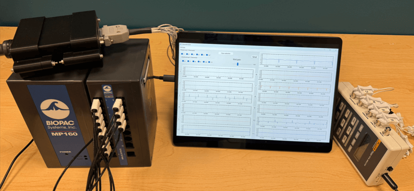
Currently conventional fetal monitoring devices are developed to focus on fetuses during labor and after 35 weeks. However, the babies who we care for at Children’s National Hospital often need medical therapy much early in gestation. This new device has been tested as early as 18 weeks with success at seeing the fetal heart signal. Dr. Govindan’s signal processing team has also reduced the need for signal averaging (averaging hundreds or thousands of beats together). The research team ultimately wants to be able to detect every individual heartbeat for treating arrhythmias which cannot be achieved using signal averaging.
This new device is a breakthrough for care of babies before birth. EKGs are one of the most used tests in medicine. Dr. Krishnan and her team believe that once the device is finalized, it will have widespread applications well beyond just treating arrhythmias. Potentially, this new device can be used to make fetal cardiac MRI easier, to carefully monitor for safety while mothers are receiving treatment with medications, to look for damage from viruses and other conditions and to prevent stillbirth in high-risk pregnancies.
Who should partner with us?
Dr. Krishnan is interested in partnering with clinicians with new ideas for innovative uses, academic centers interested in participating in clinical research testing and teams or companies who are innovative in the fields of sensors and electrodes. This project has been supported through federal grants and philanthropy. The fetal EKG research team are currently actively pursuing the next stages of funding through multiple mechanisms.
Related News
-
Bringing advanced imaging to patients around the world
June 25, 2026 -
A multisite study identifies early differences in brain development among newborns exposed to opioids before birth
June 04, 2026 -
Children’s National and Virginia Tech deepen pediatric AI collaboration to accelerate innovation for children
June 02, 2026