Our Research
Taxonomies:
Articles are not null: True
Article Count: 0
Error:
Publish Time: 12/29/2024 5:58:40 AM
-
Our studies are uncovering the role of several cellular compartments, calcium-binding proteins and lipids in cell repair. These include the role of lysosomes, which are the major calcium-triggered secretory compartments in mammalian cells. Defect in fusion of lysosomes impairs release of the lipid-modifying lysosomal enzyme, acid sphingomyelinase (ASM), and results in diseases such as dysferlinopathy or limb-girdle muscular dystrophy 2B (LGMD2B). Fusion of lysosomes and other calcium-regulated organelles in cells is controlled by multiple calcium-binding proteins, including annexins, dysferlin, and synaptotagmins, that we have examined for their role in calcium-triggered vesicle fusion during cell repair. Our work has also identified the need for the cytosol and endoplasmic reticulum to buffer injury-triggered calcium increase to control cell repair. A deficit in this process contributes to limb-girdle muscular dystrophy 2B (LGMD2L). Additionally, we find that beyond triggering membrane fusion, calcium also triggers membrane scission by the endosomal sorting complex required for transport (ESCRT) machinery, which is required for cell repair. This is regulated by the calcium-brining protein Apoptosis linked gene-2 (ALG-2) to direct scission and shedding of the damaged parts of the plasma membrane to help close a wound. We are continuing to study additional regulators of calcium-dependent membrane trafficking and their role in cell repair.
Further reading:- Dysferlin regulates cell membrane repair by facilitating injury-triggered acid sphingomyelinase secretion
- Dysregulated calcium homeostasis prevents plasma membrane repair in Anoctamin 5/TMEM16E-deficient patient muscle cells
- Injured astrocytes are repaired by Synaptotagmin XI-regulated lysosome exocytosis
- Mechanism of Ca2+-triggered ESCRT assembly and regulation of cell membrane repair
- Early Endosomes Undergo Calcium-Triggered Exocytosis and Enable Repair of Diffuse and Focal Plasma Membrane Injury
-
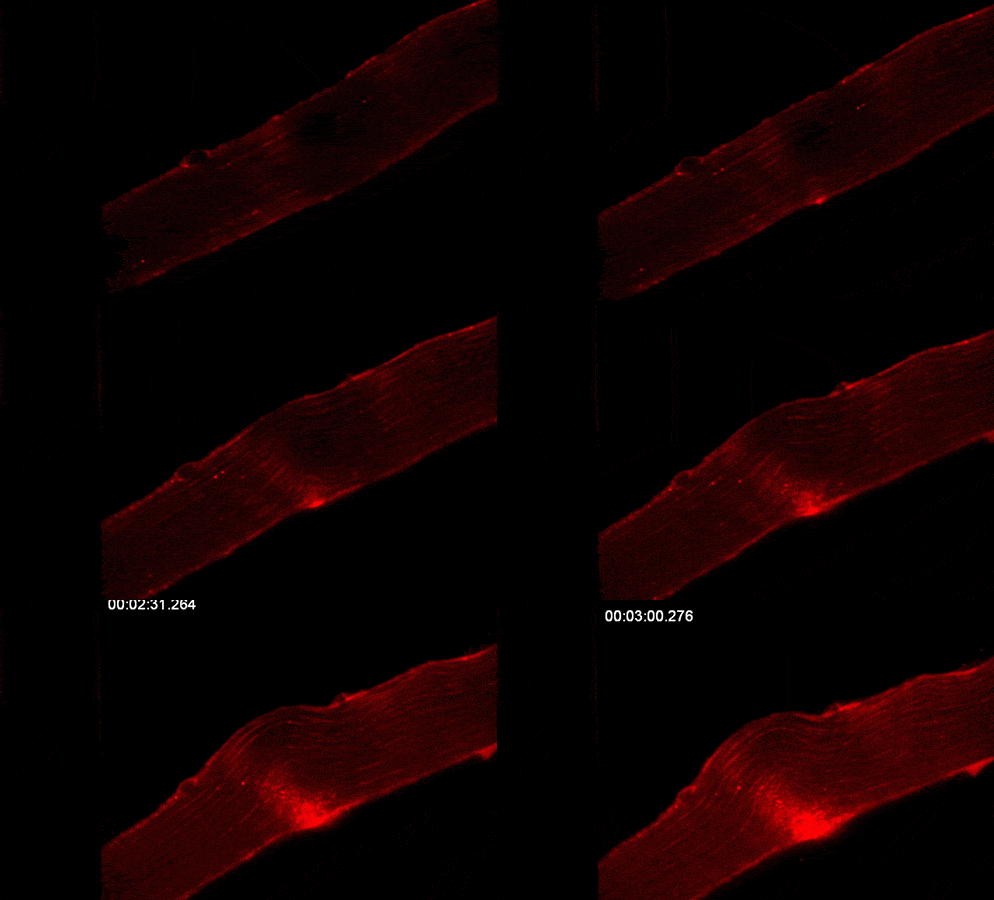
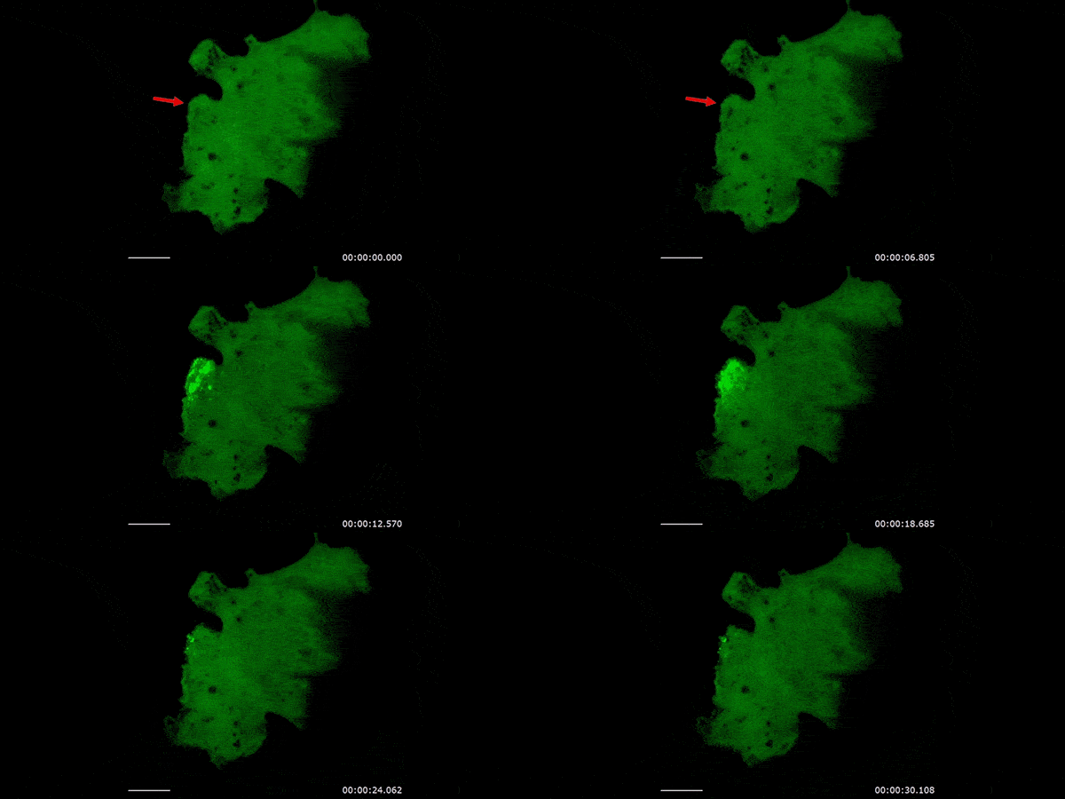
A serendipitous discovery that mitochondrial proteins accumulate at the injured cell membrane and mitochondria in muscle fiber can accumulate at the site of injury led us to studies that have uncovered the requirement of mitochondria in repairing cell membrane injury. We find that calcium that enters the injured cell is taken up by mitochondria in a regulated manner which allows controlled production of reactive oxygen species (ROS). This ROS locally activates the reorganization of actin cytoskeleton to enable closure of the cellular wound. Despite being organized in a cell-wide network, mitochondria can act locally by controlled mitochondrial fission at the site of injury. We study how mutations that alter mitochondrial calcium uptake, ROS production or fission compromise cell repair and lead to muscle diseases. Injury-mediated regulation of these processes in healthy cells, their downstream effectors, and the role of mitochondrial interactions with other organelles in controlling this repair mechanism are some of the other open questions.
Further reading:- Mitochondrial redox signaling enables repair of injured skeletal muscle cells
- Mitochondria mediate cell membrane repair and contribute to Duchenne muscular
- Use of Quantitative Membrane Proteomics Identifies a Novel Role of Mitochondria in Healing Injured Muscles
- Mitochondrial fragmentation enables localized signaling required for cell repair
-
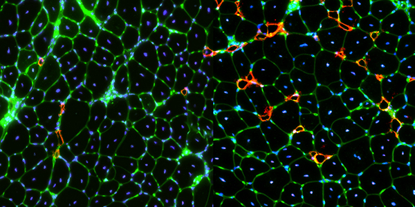
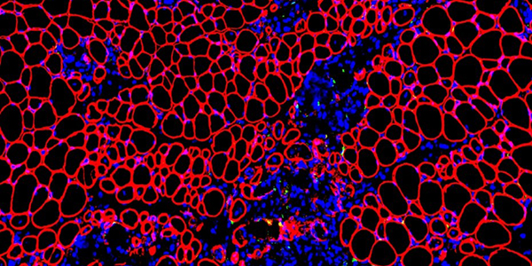
While skeletal muscle primarily consists of syncytial cells called myofibers, interactions between several muscle-resident mononucleated cell types enable skeletal muscle maintenance and repair. Mesenchymal stem cells called fibroadipogenic progenitors (FAPs) constitute one such cell type. FAPs enable homeostatic maintenance of skeletal muscle and are required for muscle repair. However, dysregulation of FAP leads to failed repair and degeneration of muscles in multiple muscular dystrophies. Using models for LGMD2B and Duchenne muscular dystrophy (DMD), we find that extracellular signaling by proteins including Annexin A2 and TGFβ cause the FAPs to accumulate and undergo osteogenic, fibrotic or adipogenic differentiation in a disease-specific manner. We are examining how FAPs facilitate repair of healthy muscles, and how altered extracellular environment in diseased muscle alters proliferation and differentiation FAPs, inhibits myogenesis and contributes to the progressive muscle loss that marks these diseases.
Further reading:- Fibroadipogenic progenitors are responsible for muscle loss in limb girdle muscular dystrophy 2B
- TGF-β–driven muscle degeneration and failed regeneration underlie disease onset in a DMD mouse model
- Contribution of Extracellular Vesicles in Rebuilding Injured Muscles
- Single-cell transcriptomic analysis of the identity and function of fibro/adipogenic progenitors in healthy and dystrophic muscle
- Pathogenic role and therapeutic potential of fibro-adipogenic progenitors in muscle disease
-
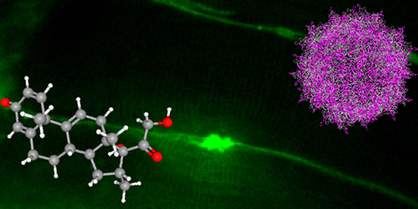
Several genetic and metabolic diseases interfere with the efficient repair capacity of the muscle, leading to debilitating neuromuscular diseases. These include limb-girdle muscular dystrophies, Duchenne muscular dystrophy (DMD) and multiple myopathies. Extending our basic discoveries, we are involved in developing and testing drugs to treat diseases involving muscle injury and degeneration. These studies contributed to the development of Vamorolone, a modified steroid analog that efficiently stabilizes cell membranes, reducing their susceptibility to injury and improving their ability to repair. It is also a potent anti-inflammatory agent, which helps reduce chronic inflammation of diseased muscles. This drug has shown safety and efficacy in preclinical studies for LGMD2B and human trials for DMD. In an alternate approach, we are employing adeno associated virus (AAV) vector for a gene therapy approach to address reduced ASM secretion by LGMD2B muscle. In another collaborative gene therapy approach, we are working to enhance the efficacy of antisense oligonucleotide-based drug delivery for treating DMD. We are also testing drugs that target FAPs to reduce muscle loss in muscular dystrophies. Another collaborative study established the benefit of low-intensity exercise to address symptoms and muscle damage in myositis patients by improving mitochondrial function. These multipronged studies are beginning to translate our discoveries to target muscle diseases that result from poor repair of injured muscle cells or tissues.
Further reading:- Membrane Stabilization by Modified Steroid Offers a Potential Therapy for Muscular Dystrophy Due to Dysferlin Deficit
- VBP15, a novel anti‐inflammatory and membrane‐stabilizer, improves muscular dystrophy without side effects
- Myoblasts and macrophages are required for therapeutic morpholino antisense oligonucleotide delivery to dystrophic muscle
- Mitochondrial dysfunction and role of harakiri in the pathogenesis of myositis
- Secreted acid sphingomyelinase as a potential gene therapy for limb girdle muscular dystrophy 2B
-
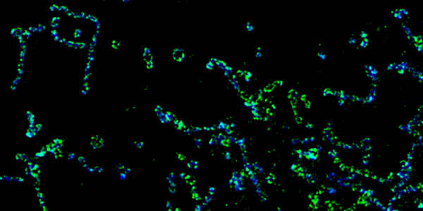
Viruses tap into the inner workings of the cell to support their own proliferation and spread, revealing many hidden workings of the cell. Aside from giving insights into cellular machinery, an understanding of these mechanisms can guide safe antiviral therapies. Through collaborations with virologists and clinicians, we study host-cell interactions with viruses including human cytomegalovirus (HCMV), human immunodeficiency virus (HIV), respiratory syncytial virus (RSV), and most recently, SARS-CoV-2. These studies have led to insights into HIV-1 entry into the renal epithelium, and the subversion of endoplasmic reticulum (ER) mitochondria contacts by HCMV. HCMV organizes its proteins in a nanoscale compartment at these contacts to prevent apoptotic death of the infected cell and uses these contacts to transport proteins from the ER to mitochondria. Beyond the cell, viruses such as RSV and SARS-Cov-2 also subvert the immune response to support their entry and prevent clearance by the immune system. We are studying the systemic responses by the airway epithelium to these respiratory viruses and how genetic and other factors disrupt these responses.
Further reading:- Superresolution Imaging Identifies That Conventional Trafficking Pathways Are Not Essential for Endoplasmic Reticulum to Outer Mitochondrial Membrane Protein Transport
- Superresolution imaging of viral protein trafficking
- Superresolution Imaging of Human Cytomegalovirus vMIA Localization in Sub-Mitochondrial Compartments
- Transmembrane TNF-a Facilitates HIV-1 Infection of Podocytes Cultured from Children with HIV-Associated Nephropathy
- IFN Stimulates ACE2 Expression in Pediatric Airway Epithelial Cells